Introduction
Complete failure of free tissue transfer is now an uncommon complication due to advancements in microsurgical techniques and technology. Free flap compromise occurs most frequently in the early postoperative period, with acute reoperation rates reported of approximately 5 per cent.1,2 Neovascularisation between the recipient bed and fasciocutaneous free flaps enables independent flap perfusion, a process called ‘pedicle independence’. A study using a bovine model demonstrated that 100 per cent of free flaps were pedicle independent by postoperative day eight.3 Due to these effects on flap perfusion, free tissue transfer is typically considered robust beyond the acute postoperative phase. To date, there are no reported cases of spontaneous free flap loss occurring after this period. The aim of this paper is to present an unusual case of late spontaneous complete flap failure requiring secondary reconstruction and provide a review of the literature.
Case
A 57-year-old male presented with spontaneous progressive necrosis of a healed free flap 11 months after resection and reconstruction of a locoregionally advanced head and neck cancer. The patient had metastatic cutaneous squamous cell cancer (SCC) involving the left parotid gland, sternocleidomastoid muscle, and left level IA and IIB lymph nodes (T0N3M0). The facial nerve was clinically and radiologically involved. The index operation involved a left radical parotidectomy, mastoidectomy with planned resection of the facial nerve, partial mandibulectomy, and left level I to V modified radical neck dissection. The defect was reconstructed with an anterolateral thigh (ALT) free flap. This was anastomosed end-to-end with the left facial artery and end-to-end with a left internal jugular vein tributary, with no acute microsurgical complications. On clinical review, up to 11 months, the ALT free flap had healed well with no evidence of recurrent disease. The patient had a history of head and neck non-melanoma skin cancers and was a smoker with a 30-plus pack-year history.
Histopathology demonstrated close but clear peripheral and deep margins with evidence of perineural involvement. Subsequently, the patient underwent adjuvant radiotherapy to the ipsilateral parotid bed and neck, comprising 66 Gy delivered in 33 fractions. Additionally, curative intent radiotherapy was delivered for a right upper lobe primary lung malignancy, consisting of 48 Gy in four fractions. This synchronous primary malignancy was diagnosed on the initial work-up for the head and neck SCC. After six months, this patient developed metastatic lung disease with involvement of the right iliac crest. The patient received a protocol-driven, concurrent immunotherapy and chemotherapy (pembrolizumab, carboplatin and pemetrexed) regimen. Systemic treatment commenced eight months after the index surgery.
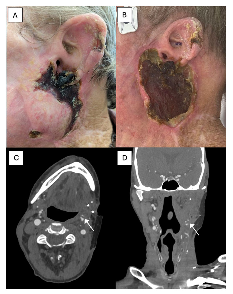
Eleven months after the primary surgery and three months into systemic therapy for the lung adenocarcinoma, the patient developed necrosis of his healed ALT free flap (Figure 1A), which progressed over the following three weeks to involve the entire ALT free flap (Figure 1B). Biopsies were negative for malignancy, and histopathological findings were consistent with acute inflammation, ulceration and necrosis. Computed tomography (CT) angiogram identified left facial artery occlusion proximal to and including the arterial anastomosis (Figure 1C and 1D). Biochemical tests and thrombotic screening did not identify a clear thromboembolic, vasculitic or autoimmune cause.
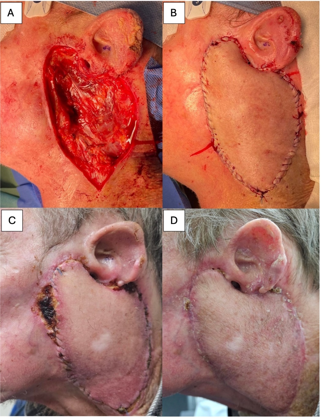
The patient underwent an exploration and debridement of the ALT free flap. Intraoperative findings identified complete necrosis of the skin, subcutaneous tissue, fascia and underlying muscle of the entire flap. There was no evidence of neovascularisation at the recipient bed. The defect was immediately reconstructed with an ipsilateral pedicled pectoralis major myocutaneous flap (Figure 2A and 2B). A pedicled flap was chosen as the preferred salvage option to avoid further potential anastomotic issues associated with a repeat free flap. Further tissue samples were negative for malignancy. Throughout this process, the patient remained systemically well with no signs of infection or disproportionate inflammation of the surrounding tissues. He actively smoked throughout.
Following the second reconstruction, the patient developed marginal necrosis of native skin surrounding the anterior flap inset three weeks postoperatively. This was managed conservatively with regular dressings and healed completely by two months (Figure 2C and 2D). The pedicled pectoralis major flap itself remained healthy throughout the postoperative course.
Discussion
This study presents a clinical case of late spontaneous complete free flap loss. For this to occur, both neovascularisation allowing for pedicle independence and perfusion through the main pedicle need to fail. Intraoperative findings during necrotic flap debridement identified negligible vascularisation of the recipient bed. The role of adjuvant radiation therapy may have been a contributing factor to the lack of neovascularisation, as the patient received adjuvant radiotherapy eight weeks after the index operation. A systematic review by Tasch and colleagues observed an increased risk of flap failure in head and neck patients receiving preoperative radiotherapy.4 Vessels exposed to radiation display wall thickening and periadventitial fibrosis, similar to vessels exhibiting atherosclerosis.5 These radiotherapy-related changes may affect the microcirculation of a flap thereby inhibiting wound healing and neovascularisation, prolonging pedicle dependency. Furthermore, there is a possibility that adjuvant radiation therapy may have caused radionecrosis to the cutaneous aspect of the flap. Acute radiation injury can result in necrosis of epithelial cells, whereas delayed necrosis occurs secondary to vascular injury and ischaemia.5 Radionecrosis and/or inhibition of neovascularisation, together with pedicle thrombosis, may have been the cause for progressive flap failure observed in this case. However, there is generally a paucity of evidence in the literature investigating the association between postoperative radiation therapy and flap failure.
A segmental pedicle thrombosis was identified on CT angiogram (Figure 1C and 1D), which is rare to occur outside the acute postoperative phase. Considering possible causes, soft tissue infections have been associated with flap necrosis in head and neck reconstruction.6 However in this case, infection was not observed and its role not established. Another cause of pedicle thrombosis could involve hypercoagulability in the setting of a concurrent malignancy. A retrospective analysis demonstrated that patients with lung cancer exhibited a higher incidence of arterial and venous thromboembolism, most pronounced in the adenocarcinoma subtype.7 This is theorised to be a result of abnormal interactions between platelet selectins and carcinoma mucins.7 Counterintuitively, while smoking is associated with adverse outcomes in terms of wound healing, current literature does not support a significant association between smoking and free flap failure in head and neck reconstruction.8
We identified a temporal relationship between starting systemic therapy and late free flap failure. In this case, the patient received a combined regimen of pembrolizumab, carboplatin and pemetrexed. There is scarce literature investigating effects of systemic immunotherapy and chemotherapy on free flap survival. One study investigating the neoadjuvant use of pembrolizumab did not demonstrate a significant association with flap failure.9 However, the association between chemotherapy and increased risk of thromboembolism is well established, particularly in patients with lung cancer treated with carboplatin.10 Carboplatin is a platinum-based chemotherapeutic and this class of agents is postulated to cause endothelial dysfunction. This may increase the risk of thromboembolism via increased cell adhesion molecules and proinflammatory processes.7 At the time of the patient’s presentation with partial flap necrosis, he had received three doses of carboplatin; this may have contributed to the arterial occlusion.
This study presents an unusual case of late spontaneous free flap failure. The generalisability of these findings are limited and should be interpreted with caution. However, this report raises the possibility of such events occurring. This is important, as it is well recognised that publication bias may result in under-reporting of undesirable patient outcomes. As a result of this clinical incident, the authors suggest that similar patients receiving adjuvant radiotherapy and platinum chemotherapies are informed during their consent process of the extremely low, although possible, risk of late flap compromise.
Conclusion
This report details a case of late spontaneous free flap failure and describes the clinical context in which this occurred. A literature review was conducted and highlights the need for further experimental and clinical research into this phenomenon. Additional data are required to better understand the complex interplay between radiotherapy, immunotherapy and chemotherapy on free flap physiology and their roles as contributors to the risk of secondary failure.
Patient consent
Patients/guardians have given informed consent to the publication of images and/or data.
Conflict of interest
The authors have no conflicts of interest to disclose.
Funding declaration
The authors received no financial support for the research, authorship, and/or publication of this article.
Revised: February 2, 2025 AEST


