Introduction
A giant cell tumour (GCT) is a rare, benign, but locally aggressive bone neoplasm that often presents late.1 Surgical management, including reconstruction, is frequently required to achieve oncological clearance due to cortical breach and soft-tissue extension, posing unique functional considerations when segmental resection is necessary.2 Metacarpal involvement presents a particular challenge in restoration of length, stability and digital alignment. We present a case of GCT of the third metacarpal, treated with wide local excision and reconstructed with a vascularised second metatarsal free flap, highlighting preoperative planning, technical considerations and postoperative recovery.
Case
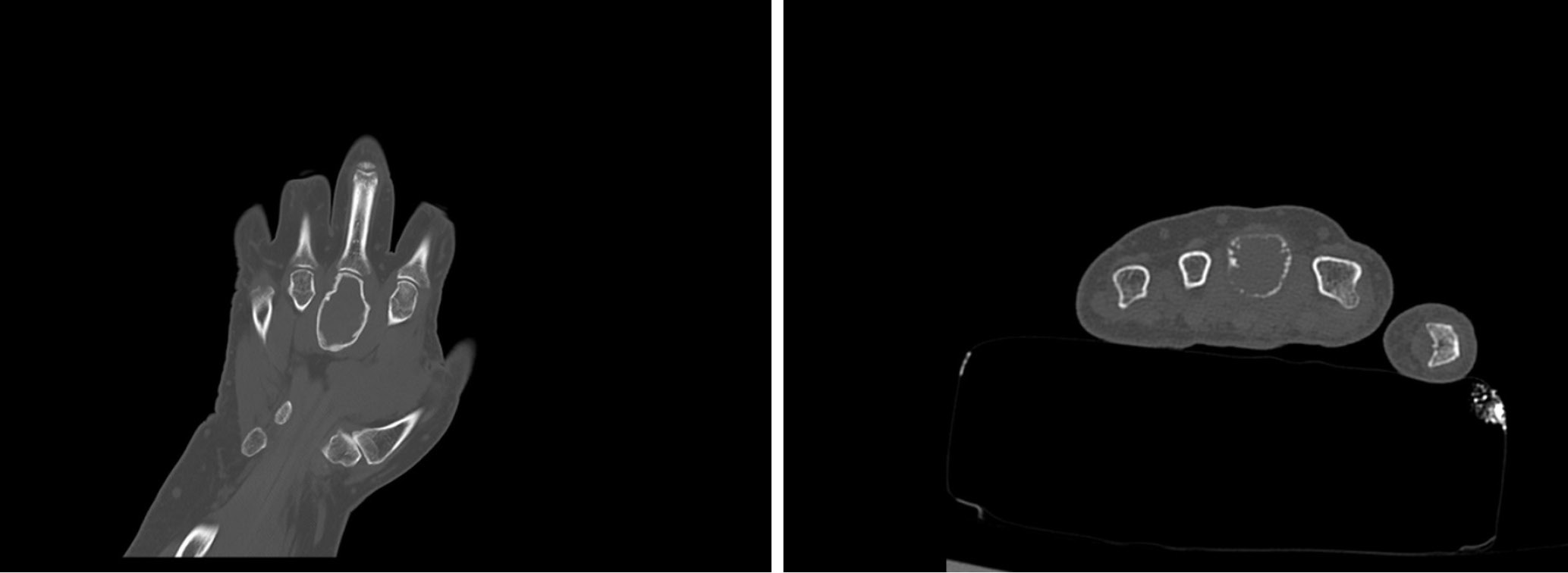
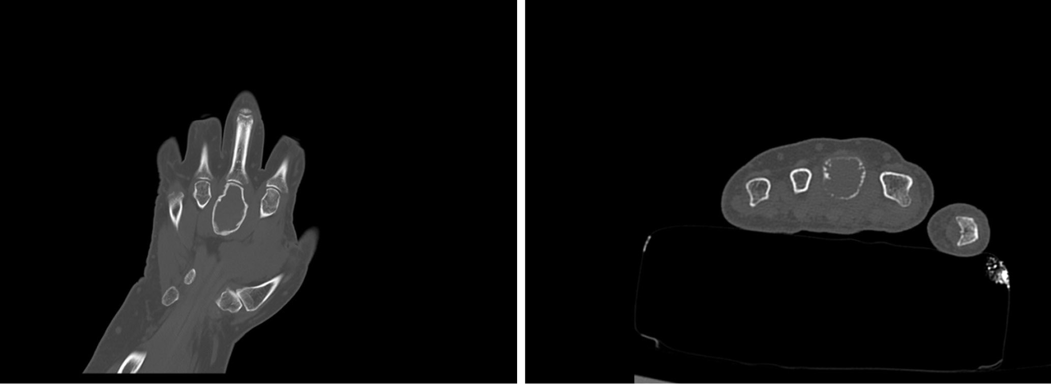
A 55-year-old woman presented with a five-month history of progressive, painful swelling on the dorsum of the left hand. Her medical history included a body mass index of 40, diabetes, hypertension, hypercholesterolaemia and non-smoker. Examination revealed a palpable mass over the third metacarpal that was radiologically investigated by preoperative CT (Figure 1), and biopsy confirmed a GCT involving the third metacarpal bone with extraperiosteal extension. Neoadjuvant denosumab was commenced and continued for three months.
It was determined, based on the extent of bony destruction, that wide local excision was required, including segmental resection of the third metacarpal bone, resulting in a 5 cm defect. A 5 cm segment of the second metatarsal of the foot was harvested to match this defect. In considering reconstructive options, a CT angiogram was conducted to determine the viability of the metatarsal flap. This investigation revealed adequate right-sided first dorsal metatarsal artery perfusion, supporting the use of a vascularised second metatarsal free flap. The risks of donor-site morbidity, healing complications and microvascular failure were discussed at length with the patient.
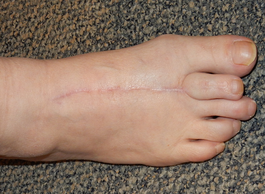
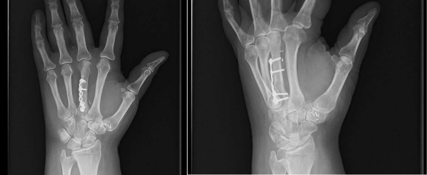
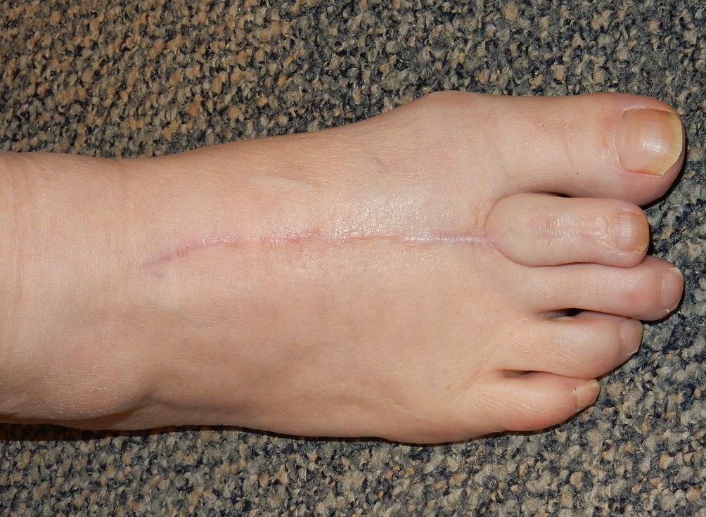
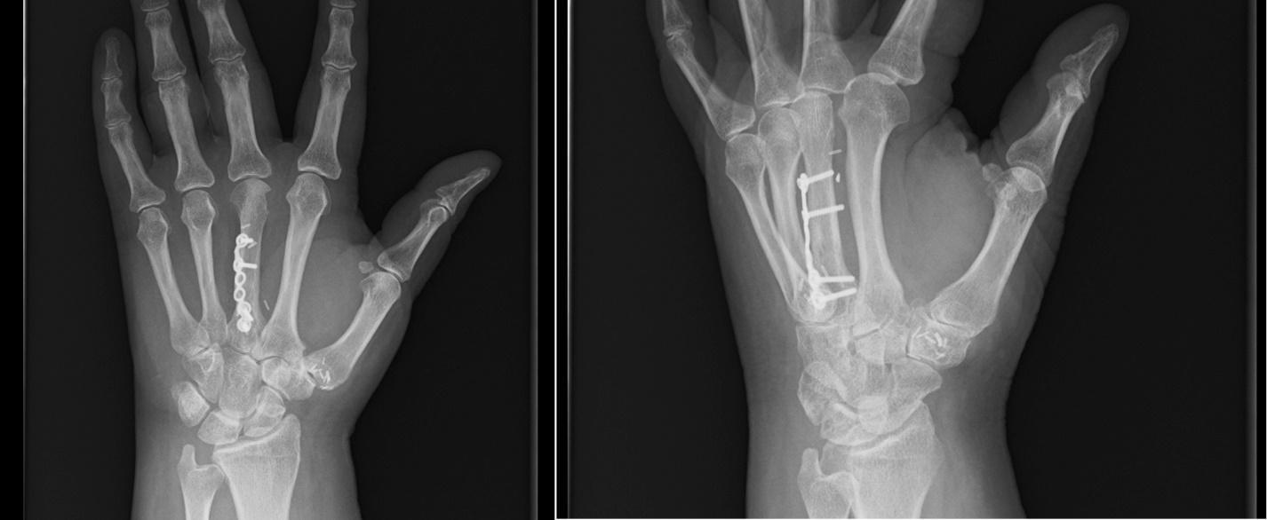
Postoperative recovery was uncomplicated, with a short period of inpatient rehabilitation. Notably, initial engagement with hand therapy was limited by prolonged swelling of the hand and emotional distress. The donor site healed satisfactorily (Figure 2). A small area of superficial skin breakdown was noted at the first postoperative dressing review and managed conservatively with non-adherent dressings. By eight weeks, the patient reported no significant foot discomfort and had returned to wearing shoes. At this review, both operative sites had healed, with improvement in digital flexion and a mild extensor lag. The nine-month review demonstrated excellent functional recovery, as shown in the Supplementary video, with full digital range of motion, stable reconstruction and no donor-site morbidity. Radiographs performed one year postoperatively (Figure 3) confirmed union and maintenance of metacarpal length and alignment.
Discussion
A GCT of the hand is rare but consistently shows more aggressive behaviour than in long bones, and treatment by curettage alone yields high rates of recurrence.3 An oncologically sound approach was therefore prioritised, with wide local excision selected as the definitive treatment strategy.
Segmental resection of the metacarpal inevitably creates several reconstructive challenges, including restoration of length and alignment, preservation of tendon balance, and maintenance of metacarpophalangeal (MCP) joint motion and stability. The literature describes a variety of reconstructive options, most commonly non-vascularised grafts from the fibula4,5 or iliac crest,6,7 and via joint-preserving osteoarticular metatarsal transfers8 or prosthetic silicone joints,9 though prosthetic joints are associated with severe problems at the MCP joint. Vascularised joint reconstructive options are reported less commonly, and are generally considered more technically demanding.10 These include vascularised fibular flaps, with or without prosthetic components, scapular bone flaps and reverse radial osteofascial flaps. Vascularised toe or metatarsophalangeal joint transfers for metacarpal GCT have been reported only rarely and predominantly for second and fourth metacarpals, with joint destruction or following trauma rather than malignancy. To our knowledge, this is the first description of a vascularised second metatarsal hemi-joint transfer to reconstruct a third MCP joint following wide local excision for GCT.
The use of a free flap in this case invites consideration of a variety of biological and biomechanical factors. The presence of diabetes and obesity presented a high risk of donor-site wound healing issues in the foot and made the flap harvest technically difficult. Poor pedal pulses on examination necessitated preoperative CT angiography, a crucial investigation rarely discussed in the context of metatarsal donor-site planning. A vascularised flap offers advantages in environments like the metacarpals, where reduced vascularity and the length of the bony defect may compromise non-vascularised bone healing and long-term joint cartilage viability. This is particularly relevant for this older patient with comorbidities, where healing would be even less reliable. The anatomy of the donor second metatarsal also provides an excellent structural match to the native metacarpal, enabling restoration of length and architecture while avoiding the need for prosthetic joint components.
Postoperatively, functional recovery was most crucial to long-term success. Expected obstacles include early oedema, stiffness and post-surgical protective splinting, but early engagement with hand therapy was essential for optimised tendon gliding, digital motion and joint stability. In this case, participation was limited by emotional distress, but the vascular nature of the flap allowed for the tolerance of delayed mobilisation without compromised union. The donor-site recovery was satisfactory, with the patient returning to wearing shoes by eight weeks and full range of motion preserved at nine months. This was consistent with previously reported outcomes of metatarsal harvest, in which long-term foot function is generally preserved. Full recovery was typically reported at 6–12 months in most case series. The decision not to restore the length of the second ray was deliberate, prioritising simplicity of donor-site closure and minimising the risk of wound complications. Given this patient’s significant comorbidities, including diabetes, obesity and poor peripheral perfusion, she was counselled preoperatively about the significant risk of donor-site complications, including skin necrosis, infection and impaired healing. Bone substitution at the donor site was therefore avoided to reduce this risk, and primary soft tissue closure was performed. While the resulting donor-site cosmesis is acknowledged as suboptimal, the patient expressed a preference to retain the second toe over ray amputation. In the absence of donor-site complications, this approach was consistent with the patient’s wishes and did not compromise functional outcomes. At nine months, the patient demonstrated preserved plantar sensation, no ulceration or pressure areas, and ambulated without a limp, with full digital range of motion, radiographic union and stable MCP joint mechanics—an excellent functional outcome consistent with results described for reconstructions using metatarsal as the donor tissue.
Conclusion
This case demonstrates that a vascularised second metatarsal hemi-joint transfer can provide a viable reconstructive option for a metacarpal GCT defect following wide local excision. It demonstrates structural and biological compatibility, minimal long-term morbidity at the donor site, and excellent functional outcomes. Given the limited literature discussing vascularised metatarsal reconstruction of oncological hand defects, especially of the metacarpal, this case adds to the evidence supporting its use in select patients. Further cases are needed to allow comparison of long-term outcomes with those of non-vascularised grafts.
Patient consent
Patients/guardians have given informed consent to the publication of images and/or data.
Conflict of interest
The authors have no conflicts of interest to disclose.
Funding declaration
The authors received no financial support for the research, authorship and/or publication of this article.
Supplementary online material
A video showing the active range of motion of the reconstructed left hand at nine months postoperatively can be found on the AJOPS YouTube channel: https://youtu.be/Vf3NqXMENBo. Please note that due to YouTube content restrictions, you may be required to sign in or create a login to access the video.