

Background
Pilomatrixomas are a benign tumour of the hair follicle.1–4 Prevalence is estimated to be less than 1 per cent of all benign skin tumours.1,3 Pilomatrixomas present as small, solitary, slow-growing subcutaneous nodules.1,3 Lesions present on the head and neck, with the face the most commonly affected.3 They are more common in adolescent years, and have a female predominance.3 Less than 10 per cent of pilomatrixomas present in patients younger than two years2 with a recent systematic review of 2189 pilomatrixomas revealing an age range of five months to 97 years.3 This case report reaffirms the need for a vigilant approach when managing paediatric skin lesions, particularly those that do not follow the expected course.
Case
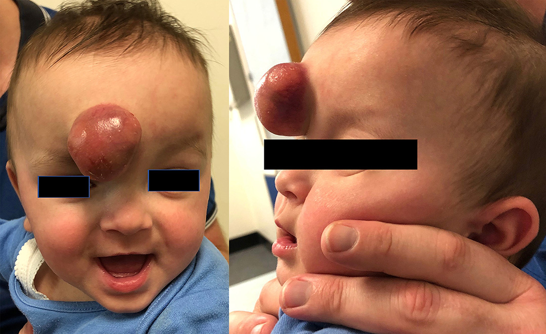
An eight-month-old boy (five months corrected), born at 27 weeks gestation, presented with a five-month history of a fast-growing, red, non- tender lesion just right of midline on the forehead. His comorbidities included prematurity, pan- hypopituitarism and an ipsilateral Horner’s syndrome from birth.
Paediatricians had initially noticed two small, red lesions on the forehead and right buttocks with the forehead lesion progressing to a large 30 × 30 × 25 mm mass by the time of referral (Figure 1). Both lesions had been diagnosed as infantile haemangiomas and propranolol had been commenced. The right buttock lesion showed signs of regression as expected, while the forehead lesion continued to grow and exhibited colour change during the subsequent months, necessitating a referral to our plastic surgery unit.

Due to the unexpected colour change and growth, the patient was sent for an ultrasound of the forehead mass, which displayed a well- circumscribed heterogenous lesion with marked vascularity, suggestive of a vascular malformation. As it was felt the ultrasound findings did not match the clinical presentation, an MRI was performed, which demonstrated a highly vascular, well-circumscribed lesion with no intra-cranial extension or osseous involvement, with features not consistent with a vascular malformation (Figure 2).

Given the atypical features, history of rapid growth and uncertain diagnosis, excisional biopsy was performed to provide clarity and exclude malignancy.
Histological examination showed a large cystic, dermally based lesion comprising basaloid cells, shadow/ghost cells and areas of focal calcification with a foreign body giant cell response, with no evidence of basaloid pleomorphism, and no atypical mitoses (Figure 3). This was consistent with a diagnosis of pilomatrixoma. The patient recovered well without further incident.

Discussion
This case is unusual for a number of reasons. The lesion was thought to resemble an infantile haemangioma, but rapid growth and colour change despite treatment questioned the diagnosis and necessitated further workup. As the ultrasound and MRI were conflicting, expert plastic surgical opinion was sought, which concluded that the lesion was unusual and warranted excisional biopsy for definitive diagnosis. The unexpected diagnosis of pilomatrixoma was made.
The diagnosis of pilomatrixomas is typically made on clinical assessment, and confirmed with histological examination after surgical excision.2,3 If diagnosis is unclear, ultrasonography can be used, which usually reveals a well-defined, hypoechoic heterogenous mass with posterior acoustic shadowing at the dermal-subcutaneous junction.2,3 Recurrence and malignant transformation are both rare.2,3
Infantile haemangiomas are more common, with an estimated prevalence of 4–5 per cent.2,5 These lesions appear early, at a median age of two weeks.2,5 They reach 80 per cent of maximal growth at five months and usually complete growth at 9–12 months.2,5 Risk factors include early gestational age and low birthweight, with prevalence up to 23 per cent in patients with a birth weight less than 1000 g.5 After a period of stabilisation, infantile haemangiomas undergo regression, with 90 per cent of cases complete by four years.2,5 After regression, 70 per cent of patients are left with a cosmetic defect such as bulky fibrofatty tissue, telangiectasia or skin laxity.5 Infantile haemangiomas are usually diagnosed clinically; however, if there is diagnostic uncertainty, imaging studies may be required.2,5 Ultrasound findings display well-circumscribed, hyper-vascular lesions with enlarged arteries and draining veins.2 Treatment options include conservative management, medical management with propranolol, surgical excision and laser therapy.3
While pilomatrixomas and infantile haemangiomas are clinically distinct entities, atypical presentation of either pathology can cause diagnostic confusion.1–3,5 Misdiagnosis can occur early when pilomatrixomas can be fairly vascular and have a blue colour. The distinction is clearer as they grow and pilomatrixomas develop their characteristic calcification. Differentiation of these two pathologies is important due to divergence in management.3 The vast majority of infantile haemangiomas can be managed expectantly, while pilomatrixoma requires excision for definitive treatment.2,3,5 When there is diagnostic uncertainty, ultrasound performed by a paediatric imaging service can usually provide diagnostic clarity; however, this was not the case in this patient and histopathological diagnosis was required.2,3,5 Atypical features of this case included a very large size, a period of rapid growth and colour change. Additionally, pilomatrixomas tend to appear in older children, whereas this presentation occurred in an infant. Ongoing rapid growth and atypical imaging features raised the suspicion of a malignant process, and a histopathological diagnosis was required.
Conclusion
We present a large pilomatrixoma presenting in an ex-premature infant. Although it is one of the most common tumours of childhood, pilomatrixomas are often not correctly diagnosed. Vigilance needs to be exercised when managing lesions that do not follow the expected clinical course and surgical excision is the appropriate treatment.
Patient consent
Patients/guardians have given informed consent to the publication of images and/or data.
Conflict of interest
The authors have no conflicts of interest to disclose.
Funding declaration
The authors received no financial support for the research, authorship and/or publication of this article.
Revised: 2021 March 5 AEST