

Background
The gracilis muscle is situated in the medial compartment of the thigh and is commonly utilised by the reconstructive surgeon due to its versatility and low donor site morbidity.1 It is a type II muscle based on Mathes and Nahai’s muscle flap classification system.2 The muscle’s dominant blood supply is from the medial circumflex femoral artery (MCFA), and minor vascular pedicles originate from the superficial femoral artery (SFA) and occasionally from the popliteal artery.3–5
The gracilis muscle may be used as a pedicled flap or for free tissue transfer to reconstruct a wide variety of defects.2,6 Popliteal defects are a complex region to reconstruct, with the literature showing overwhelming preference for local flap coverage.7,8 However, the gracilis muscle has been described for coverage of suprapatellar and anterior knee defects.9,10
This case describes the utilisation of the gracilis muscle as a free muscle transfer to reconstruct a popliteal defect. Adding to the novelty of this case, the recipient vessel used for microsurgical anastomosis was the muscle’s own minor pedicle—a technique which has not hitherto been described.
Case
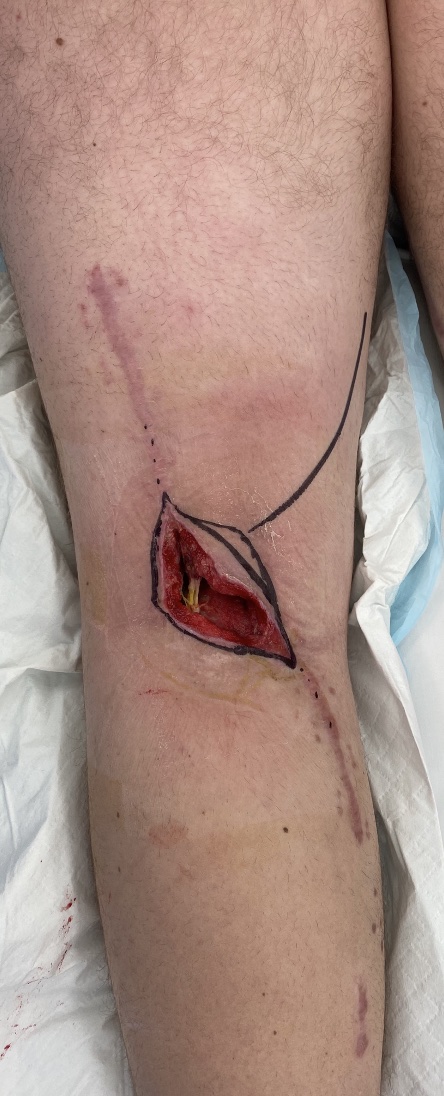
A 20-year-old male was diagnosed with a left popliteal fossa clear cell carcinoma. He underwent neoadjuvant radiation prior to aggressive resection in early 2021. Intraoperatively an 11 cm segment of popliteal artery and tibial nerve was resected and he required a saphenous vein graft and a sural nerve graft respectively for neurovascular reconstruction. The excision site was closed directly. Four months later, his wound became infected and dehisced, with nerve graft exposed. This required multiple debridements and ultimately resulted in a soft tissue defect within the irradiated popliteal fossa field (Figure 1).

Given the 11 cm segmental reconstruction of his popliteal artery, there were no suitable local reconstructive options and therefore required a free flap. An ipsilateral gracilis was raised on its major pedicle. During the flap raise, two minor pedicles were noted to be of significant calibre (approximately 2 mm diameter) (Figure 2). There were limited recipient vessels for microanastomosis due to popliteal artery reconstruction, therefore the major pedicle of the gracilis muscle was anastomosed to its most distal minor pedicle to maximise pedicle length and allow the flap to reach the defect comfortably. A split-thickness skin graft was inset over the muscle.
_gracilis_muscle_with_two_minor_pedicles_(yellow_arrows)_(b)_anastomosis_of_the_free_gr.jpg)
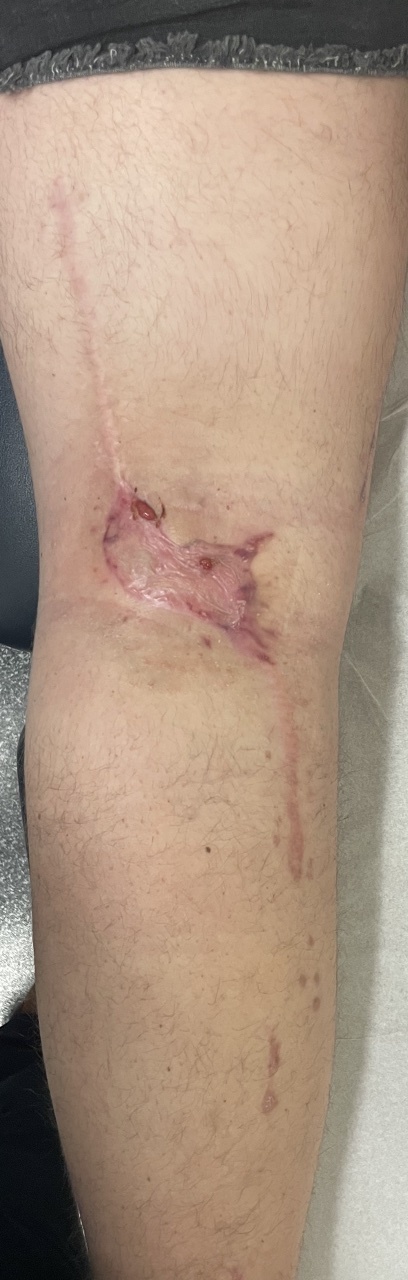
The patient recovered well postoperatively and the flap healed without complication (Figure 3).
Discussion
This illustrates the first known case of the gracilis minor pedicle used as a recipient vessel for microsurgical anastomosis. The decision to utilise this vessel was based on two factors. Firstly, there was a lack of recipient vessel options given prior resection of popliteal artery and its branches, so novel options had to be explored by necessity. Secondly, the minor pedicle of the gracilis was of good calibre, approximately 2 mm in diameter. Anastomosis of the muscle to the most distal minor pedicle allowed the flap to sit comfortably within the defect.
The anatomy of the gracilis muscle minor pedicles has been investigated extensively.3–5 It is established that there is usually at least one minor pedicle distal to the dominant supply to the muscle. This pedicle is usually from the superficial femoral artery and may be more than one vessel. Considerable variability can exist however, with minor pedicles also described as originating from the profunda femoris and obturator arteries.3
A technique described in the literature is the delay of the gracilis flap by raising the muscle on its minor pedicle and dividing its major pedicle to induce relative ischaemia and reliance on the muscle’s minor pedicle.2 This would then permit a favourable arc of rotation for a pedicled gracilis into the popliteal fossa. However, such a delay technique was not suitable in this case due to the exposed nerve graft and threatened exposure of the popliteal artery graft, which necessitated prompt coverage. There are descriptions of pedicled gracilis muscle flaps based on its minor pedicles for suprapratellar and anterior knee defects, although this requires both minor pedicles to remain intact.9,10 For more distal and posterior defects such as this case, length must be maximised and therefore this method is unsuitable.
Other studies have identified distal minor gracilis pedicles of similar calibre to our intraoperative findings of approximately 2 mm diameter.10 This is in contrast with Taylor and colleagues paper which described gracilis minor pedicles with a diameter of 1.1 mm.3 However, that paper looked at cadaveric studies, while Tiengo and colleagues paper and our findings were based on in vivo findings.10
The popliteal fossa is a difficult area for reconstruction, with limited case reports available describing strategies for soft tissue coverage.7,8 Previous examples of reconstructive options have included local reconstructive options, and regional options such as a pedicled anterolateral thigh or gastrocnemius muscle flaps.8 The gastrocnemius muscle particularly is a workhorse flap for locoregional reconstruction around the popliteal fossa and upper tibia. In our case, this was not a feasible option due to the prior segmental popliteal artery resection which sacrificed the vascular pedicle of the gastrocnemius muscle.
Conclusion
This case demonstrates an alternative novel technique for reconstruction of a popliteal fossa defect with limited locoregional options. As far as the authors are aware, this is the first case of successful anastomosis of a free gracilis muscle to its minor pedicle. Research has shown that the muscle’s minor pedicle may be of acceptable calibre in-situ for microsurgical anastomosis.7 Thus it may be considered as a recipient vessel for microsurgery in the future, especially in a similar scenario in which there has been a vascular reconstruction otherwise limiting recipient vessels. Furthermore, using the minor pedicle of the gracilis muscle as a recipient vessel is located within the region of flap elevation and may reduce operative time and donor site.
Patient consent
Patients/guardians have given informed consent to the publication of images and/or data.
Conflict of interest
The authors have no conflicts of interest to disclose.
Funding declaration
The authors received no financial support for the research, authorship, and/or publication of this article.