


Introduction
Reconstruction following burns injuries of the foot poses a difficult challenge for the plastic surgeon. The foot represents only 3.5 per cent of total body surface area. However, injuries can cause significant morbidity due to their impact on mobility.1 The need to restore both form and function requires the use of robust, pliable and thin tissue that will allow the wearing of footwear. The dearth of soft tissue in the distal foot and phalanges leads to defects that often have exposed tendon, joint or bone and may preclude the use of skin graft. In these cases, the reconstructive options are restricted to locoregional flaps, free flaps or ablative amputation. Available local flaps in the area often require the sacrifice of a major artery of the foot or potentially unreliable reverse flow. While free flaps can provide an excellent reconstruction option, they are labour intensive and have high postoperative monitoring demands. Given the known impact of amputations psychologically, and with many cultures finding amputations problematic, robust locoregional options are preferred.2,3
First described by Earley and Milner in 1989 and popularised by Quaba and colleagues in 1997, the dorsal metatarsal artery perforator (DMtAP) flap has become a useful tool in the reconstructive toolbox for defects of the foot.4,5 The operative technique is straightforward, it provides robust and thin soft tissue coverage, and the secondary defect can often be closed primarily. Given the limited number of other acceptable reconstructive options in cases with exposed joint or tendon, the use of a DMtAP flap can avoid amputation of the affected digit. Results from cadaveric studies indicate that the dorsal metatarsal artery supply is consistent, with previous studies finding over 90 per cent prevalence for the first to fourth dorsal metatarsal arteries.6–8 Cutaneous perforators are also reliably seen in all cases where the dorsal metatarsal artery is present. The clinical use of the DMtAP flap has not been widely reported, with only a handful of case series described in the literature, the most common being the first DMtAP flap.4,6–9
Operative technique
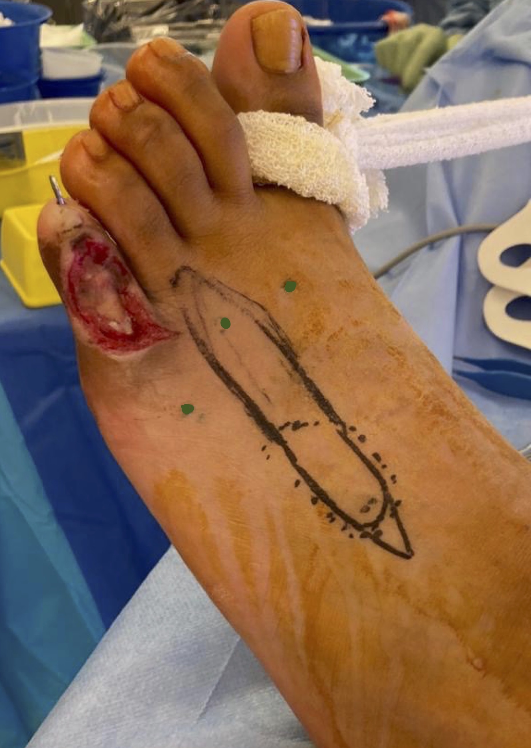
Preoperatively, the DMtAPs are mapped using Doppler ultrasound. The mapping is confirmed on the operating table using a handheld Doppler between the fourth and fifth metatarsal heads. Under general anaesthesia the patient is placed in a supine position and the defect defined (Figure 1).

If required, skeletal stability is achieved through the use of an axial K-wire. The flap is then templated to the defect, planning in reverse and with 1 cm added to ensure suitable length from the pivot point to reach the distal end of the defect (Figure 2).

The pinch test is used to ensure the secondary defect is able to be closed primarily. The flap is elevated in a subfascial plane from proximal to distal (Figure 3).
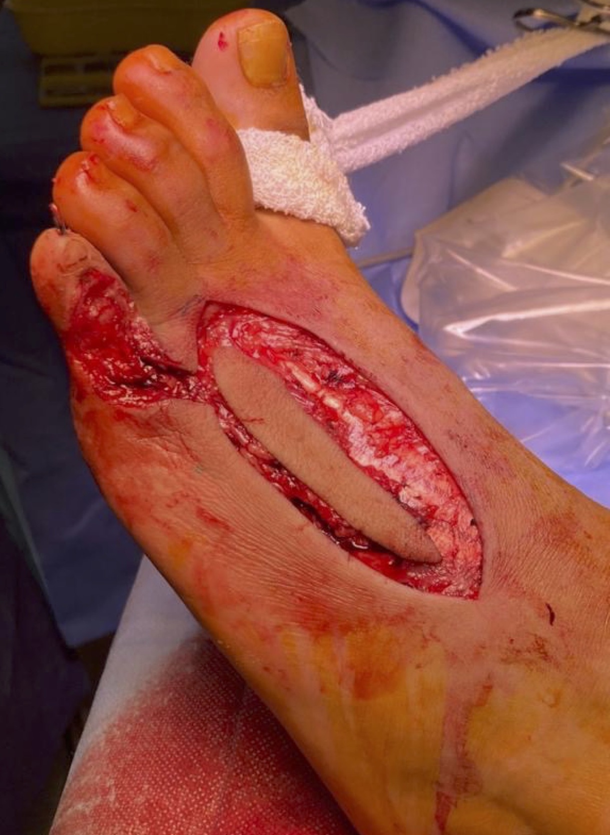
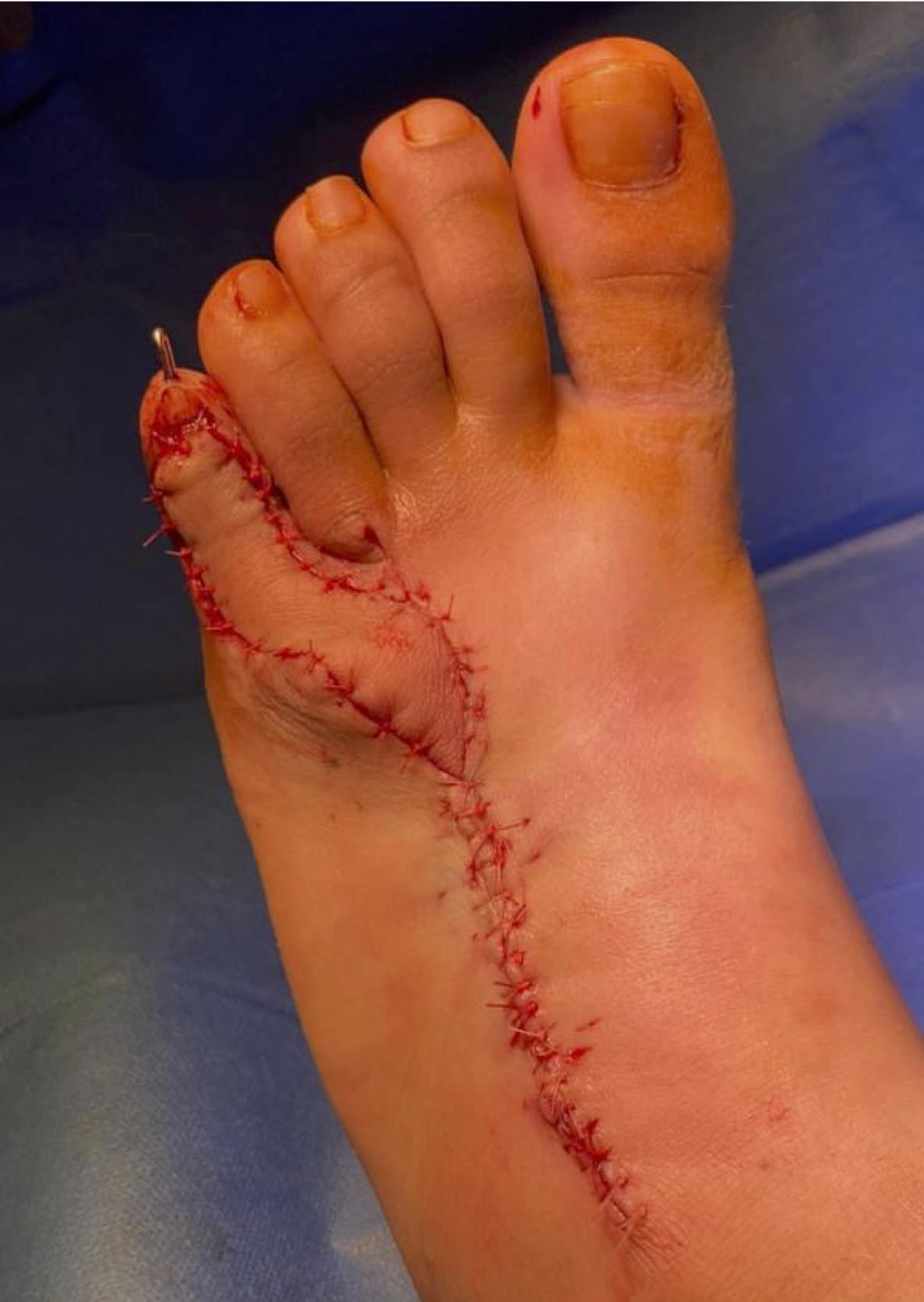
The perforator is not identified or skeletonised; the plane around the perforator is gently blunt dissected with spreading of tenotomy scissors. The tourniquet is released and haemostasis performed. The flap is then propelled 180 degrees and inset with loose interrupted sutures (Figure 4).
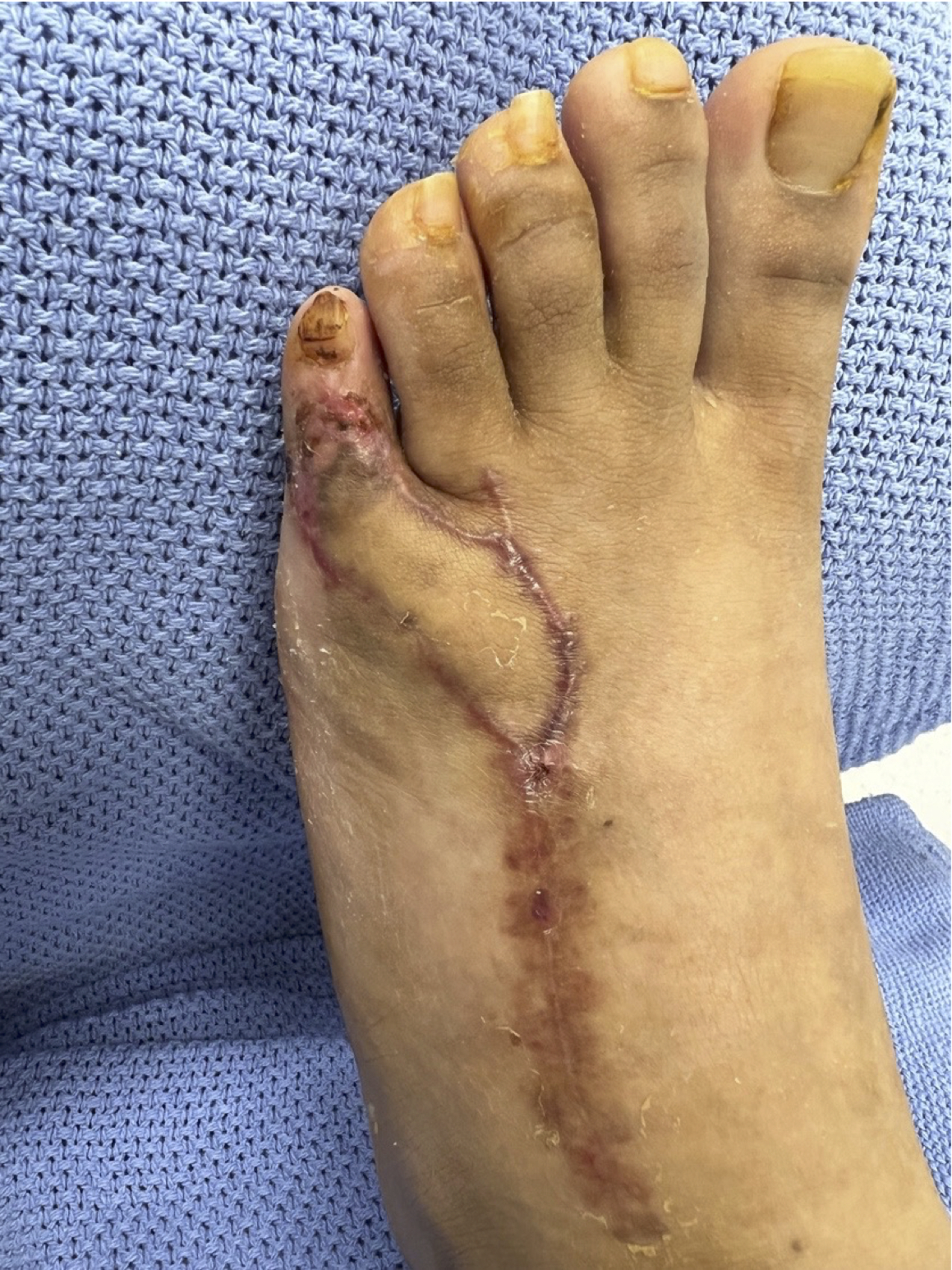
A backslab is used for stability and the leg is elevated to improve postoperative oedema. The flap is completely healed at four weeks postoperation and the K-wire is removed (Figure 5). The patient’s normal footwear is able to be reintroduced.
Discussion
Bharathwaj and Quaba, in their seminal paper, described the largest series of 14 first DMtAP flaps for burns contracture reconstruction.4 The arc of rotation for their flaps was 90 degrees; they experienced three cases of distal necrosis which healed spontaneously and suffered no complete flap loss. The donor sites in this series were grafted and the arc of rotation was 90 degrees. Hallock built on this with his series of two patients undergoing first DMtAP repair, extending the arc of rotation to 180 degrees in propeller flap style. The donor sites were closed primarily without complication.10
Cinpolat and colleagues first described the use of the third DMtAP flap and coined the term ‘metatarsal artery perforator propeller flap’.9 The authors described the use of the DMtAP flap for six patients, four with electrical burns and two with benign tumours of the forefoot. They raised five flaps based on the third DMtAP and two based on the first DMtAP, with rotation arcs ranging from 45 to 180 degrees. All flaps completely survived with no donor site complications. Van Alphen described a small fourth DMtAP flap in a six-year-old child for an open fifth toe interphalangeal joint with good functional and cosmetic outcome.7 More recently Khan and colleagues reported a third DMtAP for repair of a third webspace defect following melanoma excision.11 Taken together, the literature reports a total of 23 DMtAP flaps with a 100 per cent flap survival.
In an anatomical study, Yeo and colleagues injected 16 cadavers with red latex solution into their femoral artery.8 The limbs were then frozen for 48 hours before dissection continued under loupe magnification. A DMtAP was identified in the second to fourth webspaces in all 16 cadavers with a distal cutaneous perforator ranging in diameter from 0.5 to 0.7 mm.
Van Alphen and colleagues also conducted an anatomic study of 10 fresh cadaver feet injected with methylene blue.7 In contrast to Yeo and colleagues, they found a prevalence rate of second to fourth DMtAPs of 90 per cent. The first DMtAP was present in 80 per cent of dissections and a distal cutaneous perforator was present in all cases where there was a dorsal metatarsal artery. The fourth DMtAP was found to have the largest perforator size, with a mean diameter of 0.5 mm and a maximum perforator diameter of 0.8 mm. In this case (Figure 4), a 2.5 x 6 cm flap was able to be raised on a single distal fourth DMtAP and was propelled 180 degrees for inset.
Conclusion
The fourth DMtAP provides a reliable method for locoregional flap coverage of dorsolateral foot and fifth toe defects, potentially avoiding the need for ablative procedures. The operative technique is straightforward without the need for microsurgical techniques.
Patient consent
Patients/guardians have given informed consent to the publication of images and/or data.
Conflict of interest
The authors have no conflicts of interest to disclose.
Funding declaration
The authors received no financial support for the research, authorship and/or publication of this article.