Introduction
Since the first description by Hyde in 1883,1 the pathogenesis and clinical features of digital mucous (DM) cysts, as well as treatment options, have been well described in the literature.2 Complications associated with surgical treatment include cyst recurrence, loss of range of motion of the distal interphalangeal (DIP) joint, joint stiffness, post-surgical infection and nail dystrophies.3 Digital mucous cyst infection prior to treatment is not well documented. This report describes a complicated case of a DM cyst infection upon presentation with subsequent development of DIP joint septic arthritis, bacteremia and thoracic spinal osteomyelitis.
Case
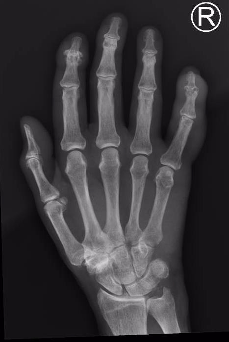
A 68-year-old female with a history of rheumatoid arthritis (not on immunosuppression) presented to hospital with a two-day history of a worsening, right little finger mucous cyst infection. She had attempted to decompress it with a needle four days earlier. Examination revealed pus from the mucous cyst with surrounding epidermolysis and evidence of proximally spreading cellulitis. She had pain on DIP joint axial loading and passive movement. A plain radiograph of her hand demonstrated swelling at the right little finger DIP joint (Figure 1).

The patient was given 1.5 g of intravenous vancomycin and urgently taken to theatre. Specialist plastic surgeons evacuated pus from the mucous cyst and the DIP joint, noting the terminal extensor tendon as ruptured but the central slip intact. Intraoperative swabs were taken and cultured, showing methicillin-sensitive Staphylococcus aureus. Blood cultures were also positive, for which infectious disease specialists suggested 1 g intravenous vancomycin twice daily. Transthoracic or transoesophageal echocardiograms found no evidence of infective endocarditis.
Daily blood cultures tested positive for eight days after admission. The patient was returned to theatre after four days as she had persistent fevers and raised inflammatory markers. Further pus was evacuated from the DIP joint, the nail removed and the nail bed and distal phalanx debrided.
Four days later, the patient was again returned to theatre where extensive epidermolysis of the little finger was debrided. The exposed extensor digitorum tendon was partly debrided and the central slip was detached and non-viable. A split thickness skin graft was harvested for wound closure and a longitudinal 0.9 mm Kirschner wire arthrodesis placed through both interphalangeal joints. Intravenous vancomycin was changed to intravenous cephazolin at 2 g three times daily, after which the patient showed no further positive blood cultures. The patient’s graft was reviewed after a further four days and considered satisfactory and she was discharged with a plan to complete another four weeks of intravenous cephazolin.
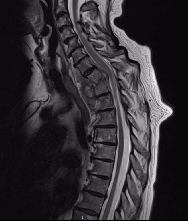
The patient presented to hospital again five weeks after discharge with severe back pain and lethargy. Computed tomography and magnetic resonance imaging revealed T1/T2 transverse myelitis/discitis with vertebral endplate destruction and a loculated paravertebral fluid collection anteriorly (Figure 2). There was mild cord compression on imaging but no symptoms or clinical signs of compression. She was recommenced intravenous cephazolin at 2 g three times daily. Her pain and inflammatory markers subsequently improved and her neurological observations were normal. She was discharged after a week in hospital with a plan for six weeks of intravenous cephazolin antibiotics. Regular outpatient reviews were conducted, including hand therapy for movement of stiff interphalangeal joints in her little finger.

Discussion
Infection is a well-documented complication following the excision of DM cysts4 but there are few reports of mucous cyst infection occurring prior to treatment. In 1984, Rangarathnam and Linscheid5 documented a series of four patients who developed infected DM cysts prior to treatment. Two of these patients described a history of trauma to the cyst, including one with an attempted needle decompression similar to the patient in this study. Bourns and Sanerkin studied a series of 20 patients with DM cysts,6 only one of which was noted to have presented with an infection after the DM cyst had spontaneously ruptured.
However, most hand surgeons would agree that infected DM cysts prior to treatment are not un- common. When they do occur, associated DIP joint pyathrosis and osteomyelitis often lead to poorer outcomes for patients, particularly in the form of joint stiffness and pain.7
Conclusion
This report, linking a mucous cyst infection with a systemic sepsis and vertebral osteomyelitis, appears to be unique. It highlights the need for aggressive management in such cases. Further study of outcome differences in DM cysts that are managed with, and without, infection would be beneficial.
Patient consent
Patients/guardians have given informed consent to the publication of images and/or data.
Disclosure
The authors have no conflicts of interest to disclose.
Funding
The authors received no financial support for the research, authorship and/or publication of this article.
Revised: November 6, 2018 AEST