Introduction
While a range of techniques for non-operative rehabilitation or surgical reconstruction of the divided or incompetent central slip have been described, the paucity of available evidence precludes direct comparison of their relative merits.1–3 A key determinant in deciding on management is whether the injury is open or closed. The presentation of patients with acute closed central slip injuries is uncommon. When these injuries do present, our institution favours full-time static splinting of the proximal interphalangeal joint (PIPJ) in full extension for six weeks. This is followed by gradual weaning of the splint and a progressive increase in PIPJ flexion for the next four weeks. By contrast, we advocate primary repair, with or without Kirschner-wiring (K-wiring), of open central slip injuries where tendon substance permits this.
Chronic closed central slip injuries, those with failed non-operative management or open injuries with loss of tendon substance all pose a challenge. Numerous surgical techniques have been described to reconstruct the central slip by either rebalancing the remaining extensor tendon or using tendon grafts.1 These techniques are not robust enough to permit early mobilisation and risk further disrupting the delicate balance of the extensor apparatus. Reconstruction with a flexor digitorum superficialis (FDS) slip, first described by Stack in 1971 using the whole FDS and subsequently modified in 2009 by Ahmad and Pickford, has been demonstrated biomechanically in cadaveric studies to provide superior strength to direct repair or lateral band centralisation.4–6 We present a case series of five patients who underwent reconstruction using a slip of FDS followed by early splintage and protected mobilisation.
Methods
Patients were selected for the prospective case series by the senior author or their senior colleagues. Suitable patients either had healed open injuries (some with loss of central slip tendon substance) or complex closed injuries with failed non-operative treatment.
Ethical approval to report these cases was obtained from the Counties Manukau Health Research Office, project approval number 861.
Case series
Five patients underwent a modified Stack reconstruction of a single-digit central slip between 2013 and 2019 (Table 1). All patients had intact intrinsic function. Only one patient, patient five, had a relevant comorbidity with a diagnosis of severe first carpometacarpal joint osteoarthritis. No wound healing or infective complications were noted.
Surgical technique
Surgery was performed by one of three senior orthopaedic hand consultants, each with over 20 years of consultant-level experience.
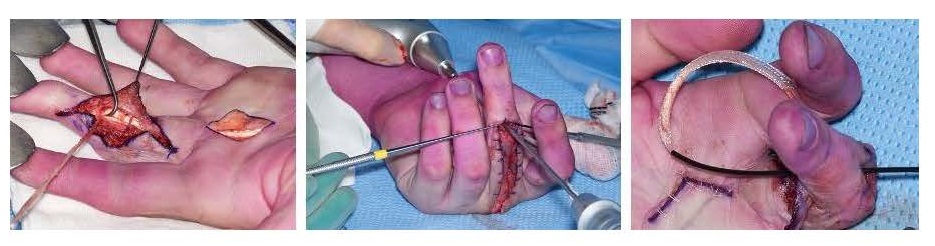
All cases were performed under general anaesthesia with an arm tourniquet. The surgical technique used was based on that described by Ahmad and Pickford with the exception that we used a volar, ulnar, hemi-Bruner incision, coupled with a midline dorsal skin incision.5 In our experience, this provides easier access than the previously described midlateral skin incision and does not result in problems of wound healing or impact upon rehabilitation. A volar, ulnar, hemi-Bruner incision at the level of A3 is used to access the ulnar slip of FDS and free this from the radial FDS slip through the chiasm. A separate oblique or chevron palmar incision is made over A1 and the ulnar slip of FDS is mobilised through both wounds and then divided proximally to produce a distally based slip (Figure 1). While Ahmad and Pickford describe using an intraosseous wire loop to split the FDS, we have not found this necessary with our more extensile skin incisions.5
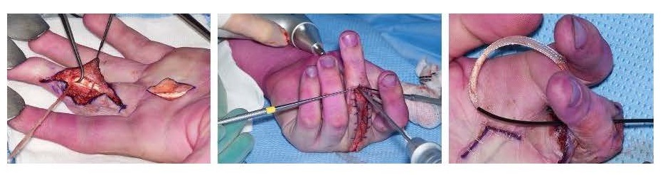
A dorsal longitudinal skin incision is used to expose the site of the central slip insertion and the zone IV extensor tendon. Under image guidance, a K-wire is inserted from the volar base of the middle phalanx to the site of the central slip insertion and over-drilled with a 2.7 mm cannulated drill. The distally based slip of FDS is passed from the volar to the dorsal incision with a tendon shuttle (QuickPass SutureLasso, Arthrex, Florida, United States) and then secured with a Pulvertaft weave into the zone IV extensor tendon. The tension is determined by tenodesis and secured with a 4–0 Ethibond suture.
Two of our patients had particular injury patterns or treatment intricacies that require further explanation.
Patient two had a missed central slip laceration following injury and a subsequent rupture one month later. At the time of listing for surgery, the patient had a full passive range of motion, but by the time they came forward for the procedure two months later, they had developed a PIPJ flexion contracture. Although we would ordinarily counsel against simultaneous joint release and tendon transfer/reconstruction, we chose to release their check rein ligaments and accessory collaterals due to the short duration of the PIPJ contracture. The patient achieved an improvement in the active extension of 90 degrees and a postoperative range of motion of 60 degrees.
Patient three had undergone K-wiring of a proximal phalanx fracture in India five years previously. Although the fracture united adequately, the patient required an extensor tenolysis in India which, regrettably, left them with no extensor function. A modified Stack reconstruction was performed after presentation to our service. This corrected their extensor lag rather too well and, subsequently, required a flexor and extensor tenolysis to achieve a satisfactory result of active extension to 20 degrees and flexion to 75 degrees with a grip strength matching that of the contralateral hand.
Postoperative management
In those patients who underwent a ‘simple’ central slip reconstruction (defined as having no other major soft tissue procedures), our default rehabilitation regime was a short arc motion protocol. This involved static splinting of the full finger for one week post-surgery. An active PIPJ flexion to 30 degrees is then allowed for one week, increasing by 10 degrees each week until postoperative week six, after which normal use is encouraged.
Outcome evaluation
Standardised range of motion measurements were recorded immediately preoperatively and during routine clinical follow-up. There was no blinding of assessors. The patients were followed up for a mean of 16 months (range 5–48 months) (see Table 2). The mean gain in PIPJ extension was 65 degrees but with a mean loss of 22 degrees of PIPJ flexion. These results occurred despite poor compliance in one patient (patient four), a simultaneous PIPJ release in one patient, and the requirement for tenolysis post-Stack procedure in another patient.
Discussion
Deficiencies of the central slip may be reconstructed by a range of techniques; some reattach the central slip or reconstruct this with a free graft while others use a lateral band to substitute for the central slip. What many of these procedures share is the need for immobilisation and lengthy rehabilitation.
Geoghegan and colleagues demonstrated that while the evidence base for both operative and non-operative treatment of acute central slip injuries is limited, the ‘evidence from individual studies tentatively supports the use of early mobilisation’.2 While it is presumptive to extrapolate such results from acute to chronic central slip management, it is not unreasonable to do so, especially if one is considering using a reconstruction of robust bone and tendon attachment. Furthermore, for a patient to regain PIPJ extension, the ability to mobilise the joint early is an important consideration in the method of reconstruction of a deficient central slip, particularly when there is a risk of losing flexion.
It was in 1971 that Stack originally described using all of the FDS to correct a Boutonnière deformity in a young builder who had two previous failed attempts at central slip repair but still retained a full passive range of PIPJ motion.4 Stack proposed that completely dividing the FDS in the palm was necessary to release a volar-deforming force at the PIPJ. He performed a tenodesis of one slip of FDS to the flexor sheath to control hyperextension. The other FDS slip, left attached at its insertion, was passed via a transosseous tunnel to the dorsum of the middle phalanx before tunnelling to the dorsum of the hand where it was tensioned and sutured to the extensor tendon in zone VI. The patient reported ‘good’ range of motion and returned to work at six weeks post-surgery.4
Stack’s procedure was subsequently modified with a single slip of FDS, via a midlateral incision, transferred to the dorsum of the finger via a transosseous tunnel and secured to the zone IV extensor mechanism via a Pulvertaft weave.5 Although no measurements were reported, they achieved a ‘functional though not full’ range of motion in a patient with established rheumatoid arthritis. This variation appealed to us due to the biomechanics of the repair and its inherent strength which affords early mobilisation. Furthermore, a recent publication reporting biomechanical cadaveric studies has confirmed that this procedure proffers a stronger reconstruction than lateral band centralisation or direct repair.6
A comparison of the results of the modified Stack technique with other reconstructions of the central slip is difficult; published studies present uncontrolled small case series, often with their own unique rating system and set of outcomes.7,8 One of the larger case series is that of Patel and colleagues who reported results of this technique in six patients, though only half had both pre- and postoperative data.7 Furthermore, two patients had closed injuries less than a month old for which we would not advocate early surgery. Nevertheless, at a mean follow-up of five months, two patients improved from an extension lag of 20 degrees and 75 degrees to 3 degrees and 15 degrees, respectively. Another patient underwent surgery in the presence of a preoperative fixed flexion deformity of 47 degrees. This improved to 40 degrees from which they could actively flex to 50 degrees. This poor result is more likely a consequence of surgery in the presence of a fixed flexion deformity rather than a failure of the actual technique.
Our patient cohort, undergoing a modified Stack reconstruction and early mobilisation, demonstrated a 65-degree mean recovery of active extension albeit at the cost of a 22-degree loss of active flexion. One patient whose finger had a complex surgical history required tenolysis following their modified Stack reconstruction. This could be considered a complication of the surgery but their complex history of two prior procedures in India possibly influenced the outcome of the modified Stack reconstruction.
Conclusion
Our series of five patients with central slip deficiencies benefit from having reliable pre- and postoperative measurements of PIPJ joint motion. From this study we conclude that a modified Stack procedure is a relatively easy technique to restore an isolated central slip function, providing a robust reconstruction that allows for early mobilisation. Our case series demonstrates an expected recovery of a 65-degree extension and a 22-degree loss of flexion. We recommend this technique as a useful addition to the hand surgeon’s armamentarium.
Acknowledgements
The senior author would like to thank Mr Hywel Dafydd, Consultant Plastic Surgeon at the Welsh Centre for Burns and Plastic Surgery, Swansea, for bringing the Stack technique to her attention.
Patient consent
Patients/guardians have given informed consent to the publication of images and/or data.
Conflict of interest
The authors have no conflicts of interest to disclose.
Funding declaration
The authors received no financial support for the research, authorship, and/or publication of this article.
The Middlemore Hospital Hand and Upper Limb Research fund receives donations via a charitable trust (Middlemore Trials). The current industrial sponsors are LMT Surgical.