Introduction
Necrotising fasciitis (NF) is an aggressive skin and soft tissue infection that can cause necrosis of the muscle fascia, subcutaneous tissues and skin.1 Streptococcus pyogenes (Group A Streptococcus, GAS), a cause of monomicrobial (type II) NF, is a highly adapted, human-restricted bacterial pathogen with a diverse clinical spectrum ranging from asymptomatic infection of the upper airway and skin through to life-threatening invasive diseases, including streptococcal toxic shock syndrome (STSS) and necrotising soft tissue infections such as NF.1
We conducted a retrospective case series of interpectoral NF caused by GAS, observed between March and July 2023—a period marked by an increased incidence of such cases internationally. The purpose of the intended case series and the details to be included were outlined to parents and informed consent was obtained as per the hospital ethics policy. Relevant patient clinical notes, and biochemical, microbiological and histological data, were obtained through a thorough interrogation of the electronic health record for each patient. Radiological reports were also obtained and reviewed for a second time by an independent radiologist. The Preferred Reporting Of Case Series in Surgery (PROCESS)2 guidelines were adhered to while investigating potential underlying mechanisms in these cases, with particular attention given to the apparent right-sided predilection of infection and the contribution of GAS.
Case 1
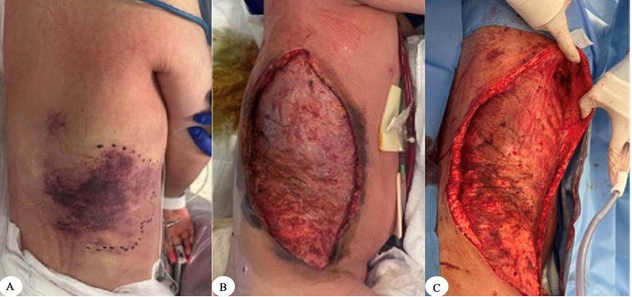
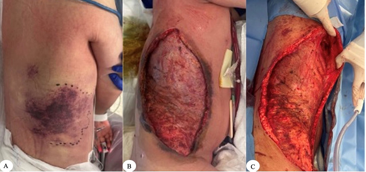
A previously well, 10-year-old boy presented to a different institution with a history of three days of fever, erythema, swelling and pain over the right chest wall. No prior trauma or other infective symptoms were reported. Clinical examination revealed pyrexia and a fluctuant swelling deep to the right pectoral muscles, with a spreading violaceous ecchymosis extending laterally from his chest to his flank and back (Figure 1). A clinical diagnosis of NF was made and broad-spectrum antibiotics were commenced (Table 1). Over hours, the patient deteriorated rapidly, becoming haemodynamically unstable from refractory septic shock, and developed multiorgan failure (MOF). Transfer of the patient to our tertiary hospital for central veno-arterial extra-corporeal membrane oxygenation (ECMO) was required, which was emergently and immediately followed by surgical debridement. Classical ‘dishwater’ fluid was encountered deep to the necrotic skin, tracking along the right interpectoral plane where matted lymph nodes and necrotic fascia were also identified. Cultures and histopathology confirmed type II GAS NF (Table 1), and antibiotic therapy was rationalised as per local guidelines. After two further debridements, the patient stabilised. The chest wall soft tissue defect was reconstructed with Novosorb (PolyNovo Biomaterials Pty Ltd) biodegradable temporising matrix (BTM) (Figure 1) and secondary split thickness skin grafting.
Case 2
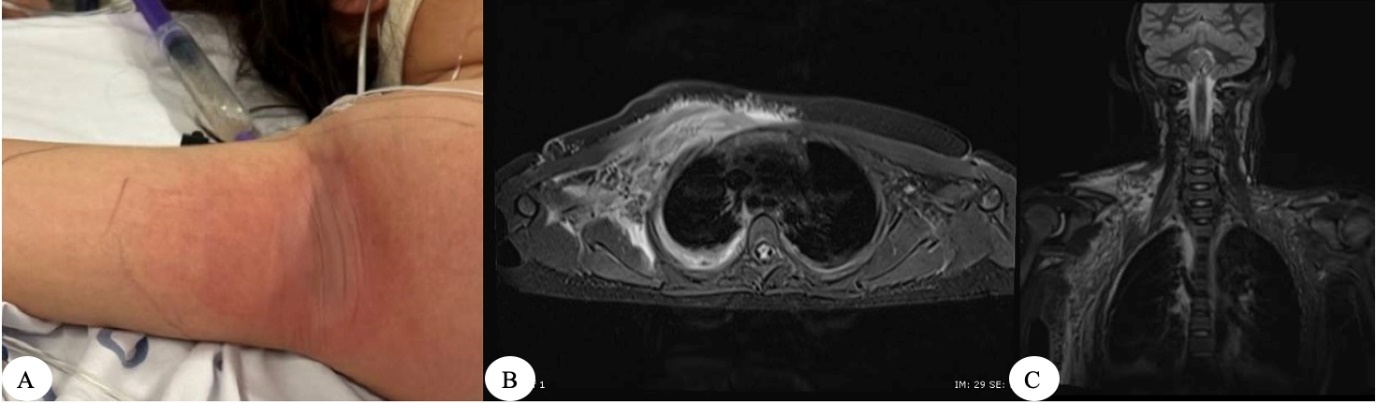
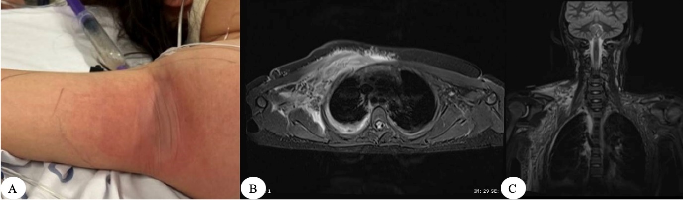
A 10-year-old previously well girl presented with a history of four days of fever and right-sided chest pain and swelling. Blanching erythema over the right chest, shoulder and axilla was noted (Figure 2). Acute deterioration with septic shock and MOF prompted initiation of broad-spectrum antibiotics (Table 1) and transfer to intensive care. Cutaneous necrosis was not present. The admitting medical team arranged CT imaging to exclude intrathoracic sepsis, and subsequent MRI following initial resuscitation to characterise the extent of soft tissue and fascial involvement. Once deeper infection was identified, the surgical team was consulted. The CT demonstrated fat-stranding in the right neck and chest, with right axillary lymphadenopathy. The MRI demonstrated subcutaneous and subfascial oedema of the right chest wall and neck, with fluid locules anterior to the right clavicle (Figure 2), but without mediastinal extension. Surgical exploration via an anterior axillary fold incision revealed necrotic pectoral fascia with characteristic dishwater fluid tracking superomedially along the interpectoral plane. Given the radiological finding of loculated fluid anterior to the clavicle, the trapezius fascia was inspected through a supraclavicular incision and found to be healthy. Fascial cultures and histology confirmed type II GAS NF; antibiotic therapy was rationalised accordingly. Two further debridements were required before physiological stabilisation was achieved. In this case, preoperative MRI did not delineate the full extent of infected tissue as reflected by the need for serial debridement.
Case 3
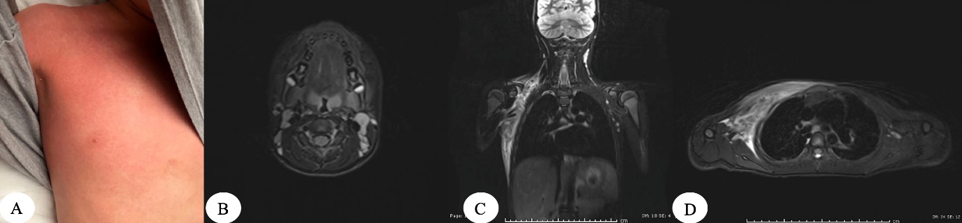
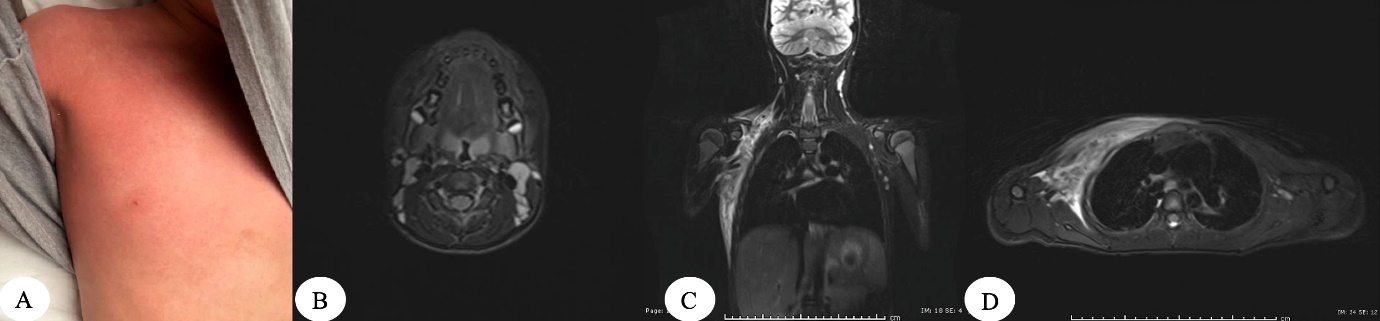
A six-year-old boy presented to primary care with a history of three days of fever, malaise and right pectoral myalgia. There was no antecedent trauma or infective illness reported. Oral antibiotics were prescribed but symptoms persisted, prompting admission under general paediatricians three days later. A fluctuant swelling under the right pectoral muscles and axillary lymphadenopathy were evident on clinical examination, with blanching erythema extending over the right chest wall, axilla and back. There was no cutaneous necrosis, bullae or surgical emphysema (Figure 3). Intravenous broad-spectrum antibiotics were started (Table 1). As the patient remained haemodynamically stable with improving inflammatory markers, an MRI was ordered to localise the deep pathology prior to surgical consultation. Subcutaneous, intramuscular and interpectoral oedema of the right chest wall and enlarged palatine tonsils were demonstrated. However, due to persistent pyrexia and progressive chest wall swelling, surgical exploration of the anterior chest wall was performed on day eight of admission. Again, the surgical findings confirmed NF in the right interpectoral plane. Necrotic fat, fascia and matted lymph nodes were debrided. Histopathological examination did not demonstrate classical features of NF; instead, inflamed granulation tissue extending into fat with neutrophils, lymphocytes, histiocytes, eosinophils and plasma cells were noted. However, the diagnosis was made on clinical and intraoperative grounds (see Table 1 for histology). Once microbiology confirmed GAS, antibiotic therapy was rationalised. The patient defervesced and recovered without further surgery.
Discussion
Necrotising fasciitis is a life-threatening skin and soft tissue infection characterised by necrosis of the muscle fascia, subcutaneous tissues and skin. Type II NF usually occurs in otherwise healthy individuals due to aggressive monomicrobial infection, commonly with GAS.3 Bacteria typically spread along the deep fascia after entry via a break in the skin caused by trauma, surgery or chronic skin conditions.4
The cases in this series bore many features typical of NF: septic shock and MOF, fish-water odour, dishwater fluid, thrombosed vessels, easily separated fascial planes, GAS within the right interpectoral plane and histologically confirmed fascial necrosis. However, none had a cutaneous portal of entry and, despite disease sufficiently advanced to cause haemodynamic collapse, two patients lacked the classic cutaneous signs of NF.
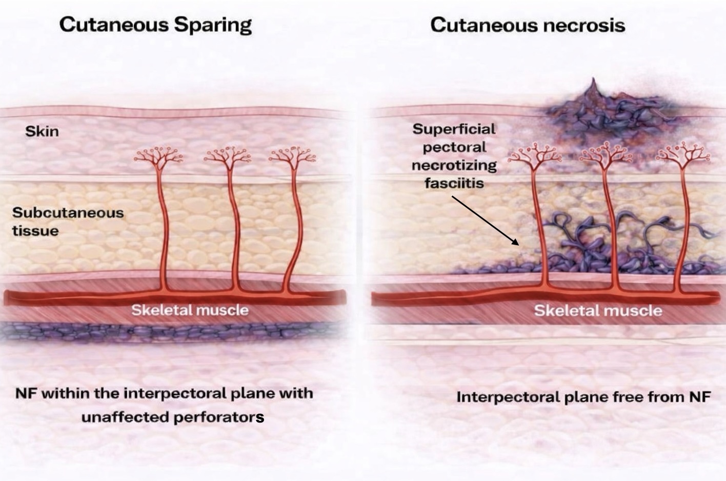
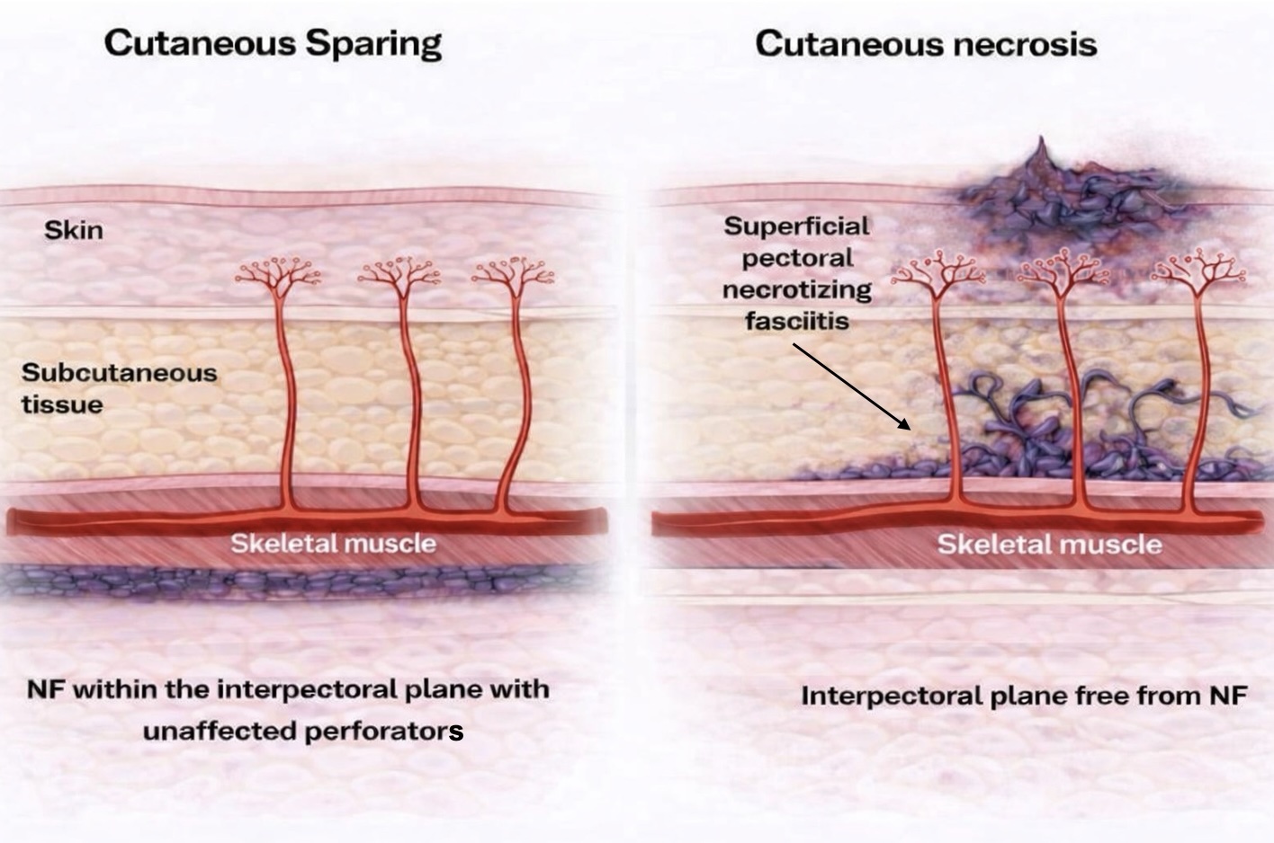
Cutaneous necrosis in NF is thought to result from microvascular compromise within infected fascial planes, including thrombosis of perforating vessels as they traverse involved superficial muscle fascia; however, necrosis is neither universal nor anatomically uniform. Angiosome theory demonstrates that thrombosis of a single perforator may have limited clinical effect due to overlapping perforasomes and dilatation of choke vessels, whereas contiguous perforator compromise may lead to cutaneous ischaemia and necrosis.5 In Cases 2 and 3, infection was confined predominantly to the deep interpectoral plane, which may explain preservation of the overlying skin despite advanced deep infection.
Large calibre thoracoacromial and lateral thoracic vessels traversing the infected interpectoral/muscle fascia presumably remained patent, as did their musculocutaneous perforators that arise superficial to the infected plane (Figure 4). In contrast, the presence of cutaneous necrosis in Case 1 may reflect thrombosis of the perforators as they traverse more superficial disease occurring along the muscle and subcutaneous tissue.
Radiological findings in this series provide an opportunity to reflect on the use of imaging in this clinical context. Cross-sectional imaging is frequently used in the assessment of suspected necrotising soft tissue infection, particularly in atypical or insidious presentations. However, it is important to emphasise that its role remains adjunctive in this condition.3,6 In this series, MRI did not reliably delineate the full extent of disease, nor did it consistently support operative decision-making, as evidenced by the need for serial debridement for one case. While MRI was helpful in confirming deep fascial involvement and excluding alternative pathology in the absence of overt cutaneous necrosis, these three cases highlight its limitations in early or atypical NF. Imaging findings should therefore be interpreted in conjunction with ongoing clinical assessment and should not be used in isolation to exclude necrotising infection.
While most associated with asymptomatic colonisation, on a global scale GAS is an important cause of morbidity and mortality. Group A Streptococcus is recognised for its ability to invade deep tissue planes and trigger systemic toxicity.7 A range of exotoxins produced by the bacterium contribute to tissue necrosis and drive the profound inflammatory response characteristic of STSS.7 In 2022, the World Health Organization reported a global spike in incidence of invasive Group A Streptococcus (iGAS).8 M1UK and related GAS lineages such as M1AUS have emerged in Australia and many other countries as a cause of severe invasive disease.9 An enhanced expression of the streptococcal pyrogenic exotoxin A (SpeA) superantigen enables the variants to evade host immunity, including in the upper airway.7 Furthermore, lymphatic vessels are an important pathway for spread of GAS infection, a process actively promoted by secreted bacterial enzymes that degrade tissue barriers.10 Lymphatic tropism in this way may explain how a subclinical upper respiratory infection led to NF in the interpectoral plane.10 An interaction between the GAS hyaluronan capsule and host lymphatic vessel endothelial receptor-1 (LVER-1) may facilitate this migration,11 overcoming the physiological barrier of unidirectional lymphatic flow maintained by intraluminal valves in some lymphatics.12 Group A Streptococcus remains extracellular during lymphatic transit,10 with bacteria moving through successive draining lymph nodes, offering further insight into possible mechanisms behind occult bacteraemia and metastatic infection encountered herein.
Radiological findings in this series provide some support for the possibility of mucosal entry: one patient had enlarged palatine tonsils; another had fat-stranding, abnormal fluid and oedema in the right neck. These findings support the hypothesis that oropharyngeal infection may extend inferiorly along contiguous or lymphatic pathways to the anterior chest wall. Similar clinical entities exist: Lemierre syndrome, a complication of bacterial pharyngitis often caused by an obligate anaerobic, gram-negative bacillus, Fusobacterium necrophorum, spreads along the lateral pharyngeal spaces and soft tissues of the neck.13,14 While Fusobacterium necrophorum provides an example of subclinical mucosal entry that spreads to cause symptomatic disease at non-contiguous metastatic sites,13,14 transmission in Lemierre syndrome occurs via vascular rather than lymphatic pathways.
All three cases in this series involved the right interpectoral plane. Given the small sample size, this laterality may be coincidental and should be interpreted with caution. Nevertheless, several anatomical and physiological factors may plausibly influence the direction of spread of infection. The complexity and variability of lymphatic drainage in the head and neck region may theoretically influence the direction of spread, although this remains speculative.15 In addition, subtle variations in local anatomy, limb dominance and exposure to trauma,16 may theoretically influence lymphatic flow, potentially facilitating localisation of infection.
Nearly half of all patients with STSS complicated by NF develop deep-seated infection at the site of minor, non-penetrating trauma or muscle strain,16 but such factors were absent in this case series. Transdiaphragmatic translocation is an established anatomical conduit for migration of infection into the thorax17; however, while right-sided thoracic involvement has been reported more frequently in some conditions, attributing the lateralisation observed in this series to this mechanism is not substantiated. Instead, radiological evidence in this study points to involvement of the mediastinum, neck and oropharynx, suggesting that the primary focus of infection may have arisen superior to the interpectoral plane and migrated inferiorly. Ultimately, however, the authors acknowledge that whether this represents a true anatomical predisposition or simple coincidence cannot be determined from a small case series of this size.
Conclusion
This case series highlights how NF can quickly progress to life-threatening severity despite a paucity of cutaneous necrosis. A high index of suspicion—guided by a thorough understanding of perforator anatomy—and prompt surgical debridement are essential to optimising patient survival and functional outcomes. This series provides insight into the invasive strategies employed by GAS and allows cautious speculation on the mechanisms underlying a possible right-sided predilection and lack of cutaneous signs observed in these paediatric patients.
Patient consent
Patients/guardians have given informed consent to the publication of images and/or data.
Conflict of interest
The authors have no conflicts of interest to disclose.
Funding declaration
The authors received no financial support for the research, authorship and/or publication of this article.